
Inspection
- Assess the patient while he/she walks back and forth on the floor and while standing. Observe step length (joint range of motion), bow-legged/knock-kneed, muscle atrophy, toeing in/toeing out.
- Consider about whether there is pelvic tilt, leg length difference, or limping. Limping is often caused by failure in the hip joint's abductor muscles. In cases of failure in the m. gluteus medius, a Trendelenburg gait can be found during clinical examination. Such a gait pattern can be caused by conditions in the hip joint (Calvé-Legg-Perthes disease, hip dysplasia, epiphysiolysis, injury and inflammation at the attachment of the abductor muscles, avulsion fracture of the greater trochanter, tendinopathy), nerve injuries that can cause failure in the abductor muscles (L5 radiculopathy), injury to the superior gluteal nerve, hip osteoarthritis, surgeries that can cause damage to the abductor muscles, hip replacement surgery, intramedullary nailing for femur fractures.
Leg length

Measurement of leg length

- Leg length is measured from the anterior superior iliac spine to the medial malleolus
- Normally 1-1.5 cm difference
Mechanical and anatomical axis
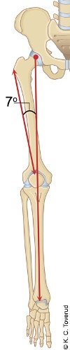
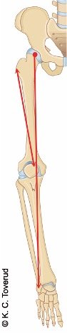
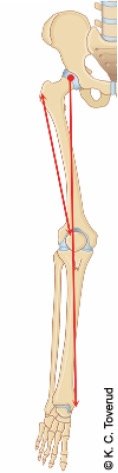
- Mechanical axis: Line from the hip joint to the ankle joint through the knee joint
- Anatomical axis: Line between the tip of the greater trochanter and the knee joint
- Angle between mechanical and anatomical axis is 7°
- Femur must tip 7° outward from the knees for the mechanical axis to pass through the femoral head
Angle between femoral neck and femur
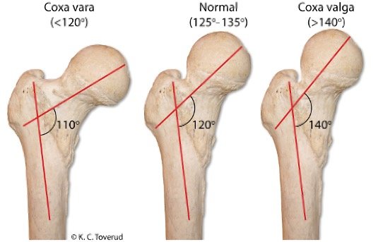
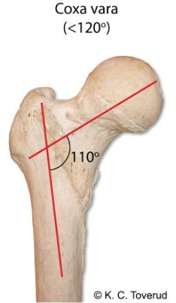
Coxa vara
Collum diaphyseal angle < 120 ˚
Can cause malalignment corresponding to the leg position in knock-knee (Genu valgum)
Normal
Angle between the femoral neck and the femoral shaft, the collum diaphyseal angle, is normally 125-135°
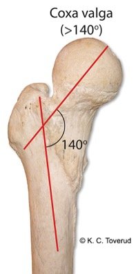
Coxa valga
Collum diaphyseal angle >140 ˚
Can cause malalignment corresponding to the leg position in bowleg (Genu varum)
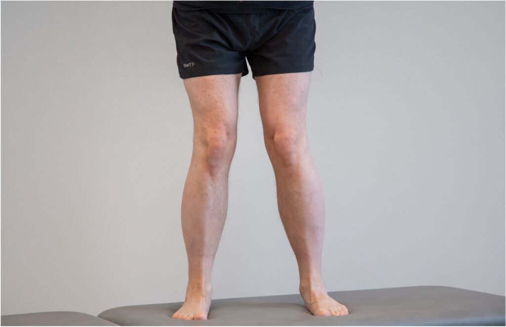
The patient is bow-legged
Genu varum
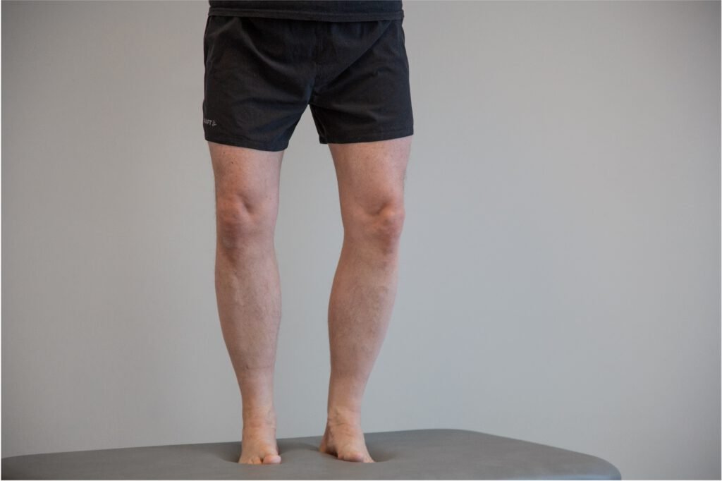
The tibiofemoral angle becomes smaller (< 7˚), coxa valga may be associated with genu varum
The patient is knock-kneed
Genu valgum
Normally, the knee joint has a bend of 5–10 degrees, called 'physiological valgus'
The tibiofemoral angle becomes larger (> 7˚), coxa vara may be associated with genu valgum
Normal anteversion angle
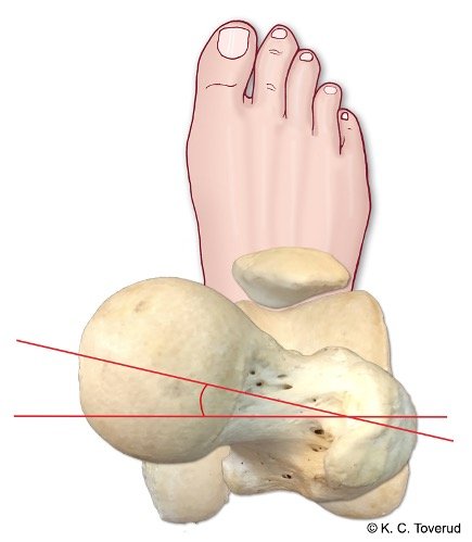
- The angle the femoral neck forms with the femoral condyle plane
- The femoral head is normally oriented 8-15 degrees forward
How to measure the anteversion angle
Normal anteversion angle


- Examining the anteversion angle in prone position
- Flexed knee joint
- Palpates that the greater trochanter is in line with the bench
Anteversion causes in-toeing
Internally rotated femoral neck, femoral anteversion


- Anteversion will lead to increased internal rotation of the femur to achieve optimal alignment between the head and the acetabulum
- Patella points too medially
- Reduced external rotation in the hips from the 0-position
Retroversion leads to outward toeing
Externally rotated femoral neck, femoral retroversion

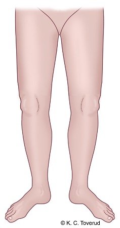
- Retroversion will result in increased external rotation of the femur to achieve an optimal fit between the head and acetabulum
- Can be rotated significantly more outward than inward from the 0-position
- Most often in men
Active movements
- Assess range of motion, pain, and side difference.
- Consider the patient's ability and willingness to move the hip joint in flexion and internal rotation → osteoarthritis?
Flexion

Supine position. The patient moves the hip joint into maximum flexion.
Extension

Prone position. The patient moves the hip joint into maximum extension.
Abduction

Side-lying position. The patient moves the hip joint into maximum abduction.
Adduction

Side-lying position. The patient moves the hip joint into maximum adduction.
Internal rotation
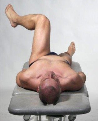
Supine position. Flexed hip joint. The patient moves the hip joint into maximum internal rotation.
External rotation
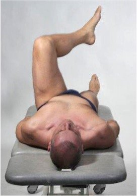
Supine position. Flexed hip joint. The patient moves the hip joint into maximum external rotation.
Passive movements
- Assess range of motion, pain, and end-feel.
- Consider whether there is a capsular movement pattern such as → Arthritis or osteoarthritis?
Flexion 120° (90-150°)
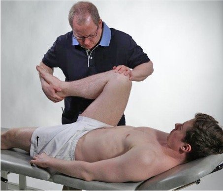
The patient is in a supine position. The examiner moves the hip joint into maximal passive flexion. One hand supports the pelvis on the opposite side to prevent rotation in the pelvis, which would give a falsely increased flexion.
Extension 10° (0-35°)
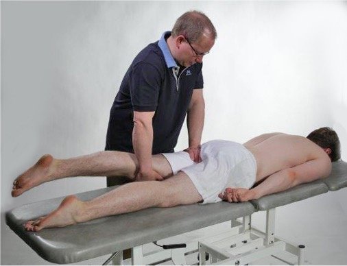
The patient is in the prone position. The examiner grasps the patient's thigh distally and supports the other hand against the hip joint posteriorly, so that the extension movement only occurs in the hip joint.
Abduction 40° (15-55°)
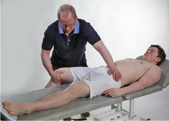
The patient is in the supine position. The examiner moves the hip joint into full abduction with the knee joint hanging passively off the table. One hand supports the pelvis on the opposite side to avoid lateral tilt of the pelvis, which would give falsely increased abduction.
Adduction 30° (15-45°)
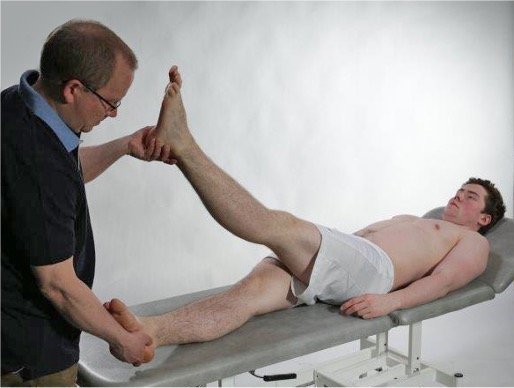
The patient is in a supine position. The examiner moves the lower limb to be examined along the bench in adduction and simultaneously lifts the other lower limb with the other hand in passive flexion.
Internal rotation 35° (20-50°)

The patient is in a prone position. Flexed knee joint. The examiner rotates both hip joints inward.
External rotation 40° (10-50°)

The patient is in a prone position. Flexed knee joint. The examiner rotates both hip joints outward.
Isometric movements
- Assess if there is muscle weakness and pain.
- Consider about the presence of power failure in combination with pain → tendinopathy, muscle rupture, and nerve involvement.
Palpation
- Assess lgroin pulse and pain over greater trochanter.
- Consider weakened groin pulse and pain over trochanter → Atherosclerosis? Trochanteric bursitis?
Groin pulse

Patient in supine position. Identify a. femoralis and assess pulse in both groins.
A. femoralis
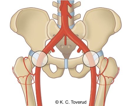
The femoral artery is found by drawing a line from the anterior superior iliac spine to the symphysis. Palpate the artery at a point on the line 1/3 of the distance from the symphysis.
Greater trochanter
The greater trochanter serves as the attachment for the gluteus maximus, medius, minimus, and tensor fasciae latae muscles, and passes just above the greater trochanter before forming the iliotibial tract. When the muscle is tight, it presses on the underlying trochanteric bursa. Increased pressure on the bursa from a tight overlying muscle can lead to bursitis. The gluteus medius prevents adduction at the hip and is the main stabilizer of the pelvis.
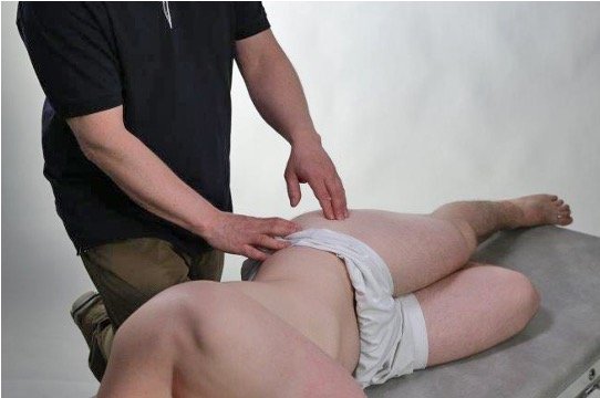
The patient is in the lateral position. Palpate the trochanteric bursa on the top of the greater trochanter. Then identify the top of the greater trochanter and palpate the attachment for m. gluteus medius and minimus. Pain located laterally in the hip joint can in this way be reproduced by palpation over the trochanter area and by using the FABER test.
M. gluteus medius
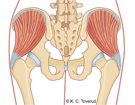
M. gluteus medius with attachment on the greater trochanter. Degenerative changes in the tendon attachment can lead to muscle atrophy and weakness. This can lead to an unstable pelvis.
Pain area in tendinopathy of m. gluteus medius

Trochanteric tendinopathy/bursitis

The cause of tendinopathy in the gluteus medius muscle is degenerative changes with small partial ruptures in the tendon at its attachment on the top of the trochanter. Leads to atrophy in the muscle with subsequent pelvic instability.
Trochanteric bursitis is assumed to be a consequence of the tendinopathy and is caused by increased compression from the tensor fascia lata over the bursa in an unstable pelvis.
Assessment of clinical presentation based on functional examination
The most common condition in middle-aged and elderly people is hip osteoarthritis. The diagnosis is often clear from the medical history, pain at the start of movement, and limping gait. In the clinical examination of hip osteoarthritis, one often finds a capsular pattern of movement with reduced flexion and internal rotation in the early stages of osteoarthritis development; sometimes the clinical pattern is present before it is possible to see osteoarthritic changes on skeletal X-rays.
If there is a non-capsular movement pattern, one must consider prosthetic solution or bursitis. Bursitis in the hip region can be located over the trochanters. You can then provoke pain with passive abduction and by palpation over the greater trochanter. In some cases, the bursitis may be located under the psoas muscle at the surgical neck. You can then find pain in the groin with passive flexion in the hip joint in combination with passive adduction. In some cases, there may be so-called gluteal bursitis, most often located in the layer between the gluteus minimus and medius muscles. The diagnosis can often be difficult to establish; patients often report pain over the gluteal area with passive flexion. in the hip joint. In addition, patients may experience pain provocation during isometric internal rotation and/or during isometric external rotation in the hip joint, probably due to compression of the bursa between the muscle bellies. On palpation, it may be difficult to distinguish between gluteal bursitis and myalgia in the gluteus medius muscle; the latter condition is a very common condition that can cause pain in the lateral hip region.
During examination of isometric flexion in the hip joint, pain may be triggered in younger individuals with a history of activity-related hip/groin pain as a sign that a psoas tendinopathy may be present. With pain in the groin towards the symphysis, one may suspect that there could be an inflammation at the attachment of the m. adductor longus; patients with this issue will often respond with pain during isometric adduction in the hip joint.
Femoroacetabular impingement (FAI) often causes activity-related groin pain and stiffness in the hip joint. Clinical examination often shows a positive impingement test and positive FABER test. There may also be a possible degenerative labral involvement, and there is a long-term risk for the development of osteoarthritis in the hip joint.
Piriformis syndrome often causes gluteal pain with radiation, worsening with sitting, and a positive provocation test.
Special tests
Final competence

- Perform relevant clinical examination in a skilled and gentle manner
- Interpret clinical findings and be able to establish tentative diagnoses
- Suggest referral and treatment
Last updated
31.08.2021