
-
Inspection
- Assess on whether there are visible abnormalities in the face, skin, and jaw function. Observe the patient both at rest and during simple movements (smile, raise eyebrows, close eyes tightly). Note any abnormalities that may indicate cranial nerve involvement, inflammation, or jaw joint problems. Document findings and assess whether further testing of cranial nerves, jaw joints, or neurological status is necessary
- Consider about the presence of discoloration, swelling, paralysis in the facial muscles, or deviation of the jaw. Consider whether any swelling or tenderness around the jaw joint, ear, or cheek is due to temporomandibular dysfunction (TMD), infection, or inflammation.

Look at the patient from the front. Note if the jaw deviates to the side when the patient has their mouth closed. -
Active movements
- Assess about the presence of normal range of motion in the jaw joint for mouth opening, lateral deviation, and protrusion. Record if the movements trigger pain and if there is asymmetry or deviation in the movement path.
- Consider whether there is reduced range of motion, pain or locking when opening the mouth. Deviation to one side may indicate muscular dysfunction or involvement of the jaw joints.
Normal opening of the mouth
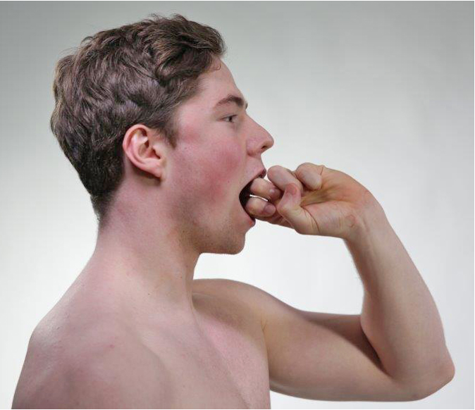
Normal opening of the mouth corresponds to 2-3 finger widths (50 mm). Lateral deviation when opening the mouth

Record whether there is a lateral deviation when opening the mouth. Lateral deviation triggers pain

Ask the patient to actively move the jaw from side to side. Record differences in range of motion between sides (normally 10 mm side movement) and whether the patient reports pain. Protrusion triggers pain
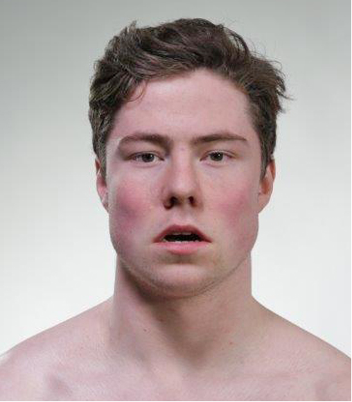
Ask the patient to push the lower jaw forward, protrusion (normally 8 mm protrusion). Record whether the patient reports pain. Good jaw function requires normal function of the neck joints and muscles in addition to the jaw joint and jaw muscles. Mouth opening is normally limited by muscles, while side movements are limited by ligaments.
(E H Julsvoll (ed.) Temporomandibular Dysfunction. Theory, Examination, Treatment. Cappelen Damm Academic 2018).
-
Passive movements
- Assess the mechanics of the jaw joint, disc function, and musculature. Stabilize the patient's head and neck before performing passive movement. Move the jaw slowly and controlled to feel for end-feel, resistance, and any clicks. Observe the patient's facial expression and ask about pain at the end point of the range of motion. Clicks may indicate disc displacement or internal dysfunction in the jaw joint. Pain during passive movement often points to joint or disc pathology, not a muscular cause.
- Consider about whether there is clicking in the joint, pain at the end point of the range of motion. Determine whether the clinical picture indicates disc problems or whether it is muscular pain (myalgia) around the jaw joint.
Lateral deviation
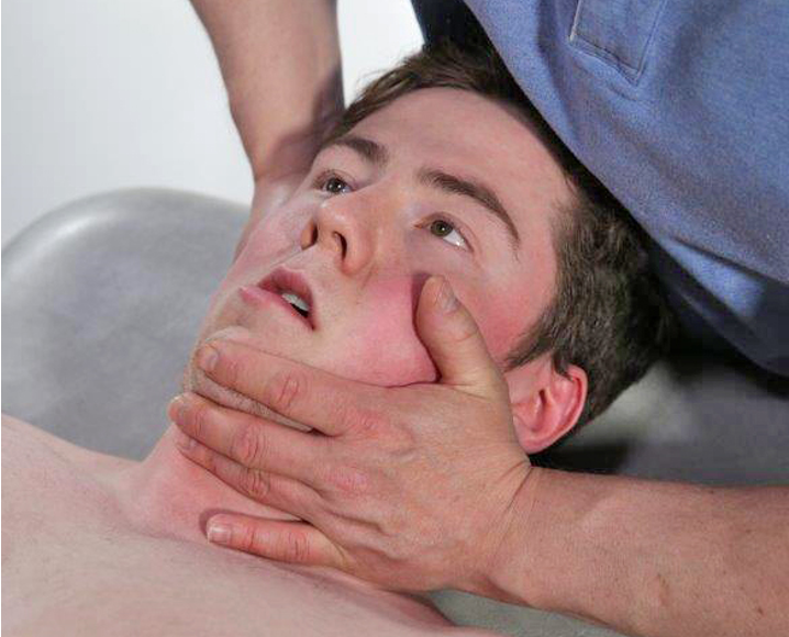
Perform a passive lateral deviation of the jaw to the right and to the left. Note clicking in the jaw joint and whether the patient reports pain at the end point of the range of motion. Clicking and pain indicate a disc problem. -
Isometric movements
- Assess Muscle strength and pain. The relevant muscles around the jaw joint are innervated by n. mandibularis (V), the motor branch of n. trigeminus.
- Consider about lateral deviation and opening against resistance triggers pain. If pain is triggered by lateral deviation, the cause is often tendinopathy/myalgia in the pterygoid muscles or disc problems or capsulitis in the jaw joint. If pain is triggered by opening the mouth against resistance, the explanation may be tendinopathy/myalgia in the temporalis muscle or in the masseter muscle, but it can also be caused by capsulitis or disc problems.
Ask the patient to laterally deviate the jaw against resistance, record the force applied and whether the patient reports pain. Ask the patient to open the mouth against resistance, record whether there is pain. The most important muscles for lateral deviation, opening/closing the mouth, and protrusion of the jaw are the lateral and medial pterygoid muscles, masseter muscle, and temporalis muscle.
-
Palpation
- Assess on palpation of the jaw joint triggers pain, clicking, or locking. Palpate both the jaw joint and the masseter muscle systematically, with the patient in a calm and relaxed position. Examine both with the mouth closed and open. Record pain, click, crepitus, locking tendency, and any pain radiating toward the eyebrows, jaw, and area in front of the ear.
- Consider which of the clinical tests (active, passive, isometric, palpation) are positive. A clinical assessment is made as to whether the findings indicate the presence of Temporomandibular Dysfunction (TMD), muscular dysfunction, disc involvement, arthritis, or referred pain from the masseter muscle.
Palpation of the jaw joint with the mouth open
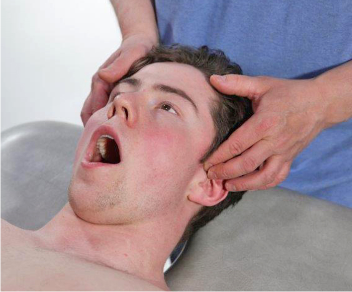
Palpation of the temporomandibular joint with the mouth closed
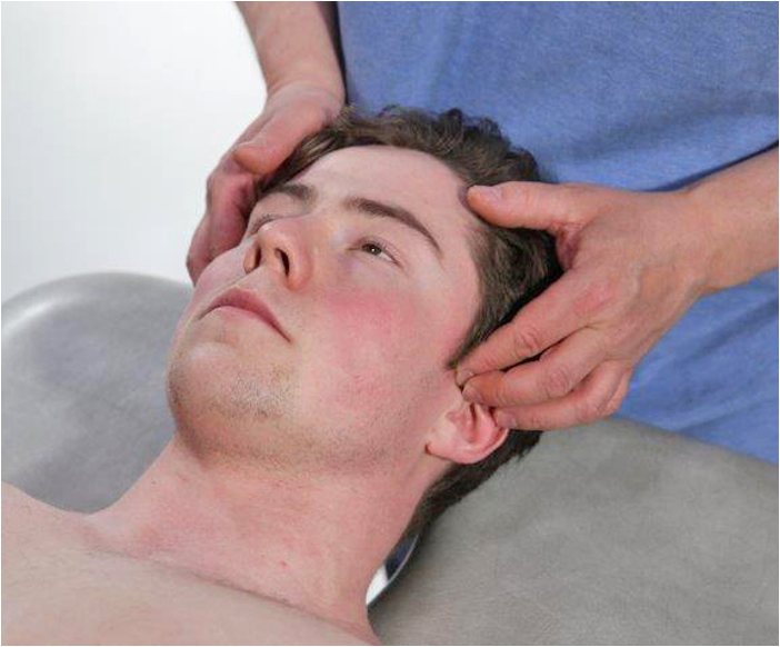
Palpate the temporomandibular joint space with the mouth open and closed. Record whether palpation of the temporomandibular joint triggers pain radiating to the ear and face, clicking, or locking tendency.
Temporomandibular Dysfunction (TMD)
The prevalence of people seeking treatment for TMD in the Norwegian population is 4-7%. The condition affects individuals aged 20-45 years, with a female-to-male ratio of 4:1. Pain is the dominant symptom.
(Norwegian Directorate of Health (2008). Treatment options for patients with temporomandibular dysfunction (TMD). Oslo: Norwegian Directorate of Health and Social Affairs. E H Julsvoll (ed.) Temporomandibular dysfunction. Theory, examination, treatment. Cappelen Damm Academic 2018).
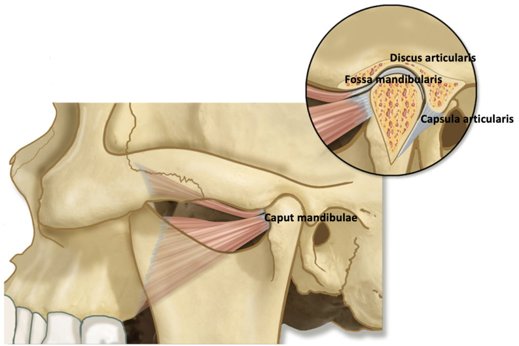
Causes of TMD
- Injury to the articular disc
- Damage to joint cartilage after arthritis
- Direct trauma to the jaw joint
Symptoms of TMD
- Pain in the face
- pain in the jaw joint and the surrounding area, including the ear
- inability to open the mouth comfortably
- clicking or creaking sounds in the jaw joint
- jaw locking when opening the mouth
- headache
- uncomfortable or uneven bite
- swelling in the area around the jaw joint
palpation of m. masseter
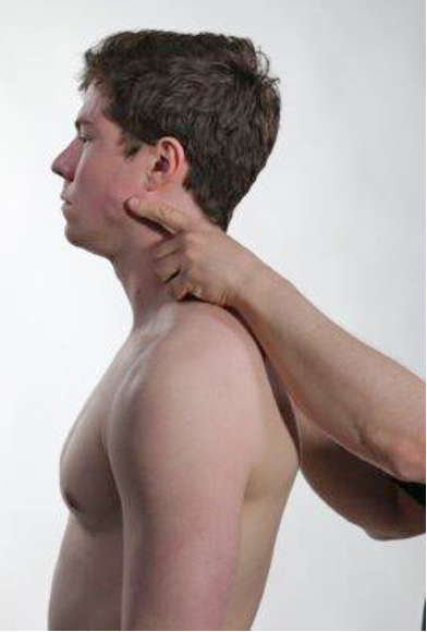
Palpate the m. masseter when the patient has their mouth open, the muscle belly is pressed against the mandible and record if palpation triggers pain radiation to the eyes, jaw joint, or towards the ear. 
Referred pain from trigger points in the m. masseter can go to the eyebrows, jaw, or ear. -
Assessment of clinical presentation based on functional examination
Temporomandibular joint pain is common; about 50-75% of the population has had at least one episode of pain in the temporomandibular joint or in the temporomandibular muscles. Most common in women over 55 years old. Pain in the temporomandibular joint and in the temporomandibular muscles is often associated with headaches. Pain in the temporomandibular joint is often most pronounced when chewing or grinding the teeth. Osteoarthritis of the temporomandibular joint can be present in older individuals. Dislocation of the disc in the temporomandibular joint is more common in somewhat younger people. Osteoarthritis and disc dislocation can lead to a tendency for temporomandibular joint locking, crepitus in the joint, and difficulty opening the mouth. Trigeminal neuralgia can cause burning pain in the temporomandibular joint area and is an important differential diagnosis for unclear cases pain in the jaw joint area. One must also be aware that sinusitis and root canal infection in the upper jaw can give referred pain to the jaw joint.
-
Special tests
-
Final competence

- Perform relevant clinical examination in a skilled and gentle manner
- Based on the medical history, set up a working hypothesis and formulate a testable problem statement
- Recognize clinical pictures/patterns of conditions in the musculoskeletal system
- Set up tentative diagnoses sorted by probability for a condition
Last updated
28.02.2022