
Inspection
- Assess to start with the patient in a standing position, relaxed posture. Observe if the patient stands tense with raised shoulders. Inspect systematically from front and back, assess symmetry, contours, muscle atrophy, and scapula position. Assess muscle bulk and bone and joint structures.
- Consider on whether there is muscle atrophy. Deltoid atrophy is a common finding in shoulder disorders due to inactivity related to the symptomatic shoulder. In rare cases, there may be axillary nerve neuropathy following an anterior shoulder dislocation. Atrophy in the infraspinatus muscle can also be assessed by inspection in the infraspinous fossa; the cause is most often compression of the suprascapular nerve in the scapular notch, or there may be a complete rupture of the muscle after trauma. Also assess whether there may be acromioclavicular joint osteoarthritis and winged scapula.
From the back
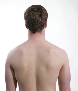
Observe whether the patient stands tense with raised shoulders and whether the scapula lies evenly against the thorax. See if there are signs of infraspinatus muscle atrophy.
From the front

Observe if there is deltoid atrophy. Look for whether the clavicle has a normal contour and if there is swelling over the acromioclavicular joint.
Active movements
- Assess the patient's ability and willingness to move the shoulder joints. Observe if movement causes pain, if there is reduced range of motion, and a movement pattern indicating a painful arc or capsulitis.
- Consider about whether there are normal ranges of motion, side differences, and whether any of the movements trigger pain → pain arc as a sign of subacromial pain syndrome (impingement). Also consider whether the pain is accompanied by weakness, which may indicate rotator cuff involvement/tear. If there is clearly restricted mobility in abduction and external rotation and you at the same time observe an early co-movement of the scapula during the abduction movement, there may be a capsulitis (frozen shoulder).
Elevation

Lift both arms up to 180° straight out to the side and completely up toward the ceiling. The movement is called elevation.
Painful arc
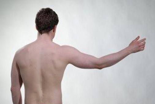
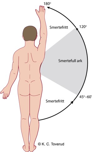
Notes on the patient indicate pain in the area between 45-60-120º as a sign of pain arc.
Subacromial pain syndrome (impingement)
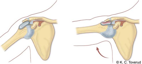
- Thickened subacromial bursa
- Calcification in the tendon attachment of m. supraspinatus, m. infraspinatus, or m. subscapularis.
- AC joint arthrosis and acromion type 2 can worsen an impingement.
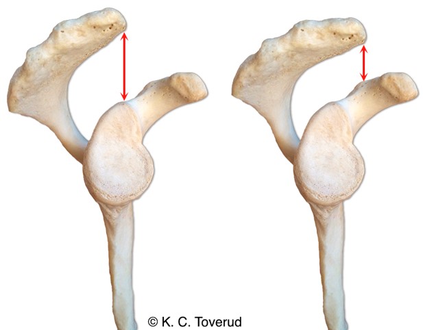
- Acromion type 1 on the left (normal) and Acromion type 2 on the right (produces narrower subacromial space)
Michener LA et al. Reliability and diagnostic accuracy of 5 physical examination tests and combination of tests for subacromial impingement. Arch Phys Med Rehabil. 2009;90(11):1898-1903
Flexion
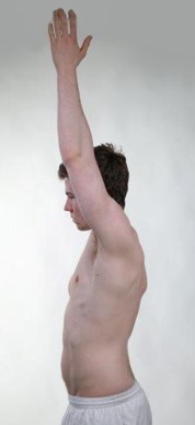
Ask the patient to lift both arms straight out in front of the body and all the way up towards the ceiling.
- Even if the patient reports pain during elevation as a sign of a painful arc, the flexion movement may be painless.
External rotation

Ask the patient to rotate both shoulders outward away from the body. Compare the ranges of motion in the right and left shoulder.
Internal rotation

Ask the patient to place one arm behind their back. Compare the ranges of motion in the right and left shoulder.
Passive movements
- Assess about whether the ranges of motion are normal, whether they trigger pain, and what end-feel is present. Examine passive shoulder movement systematically, starting with elevation, then perform abduction with the scapula fixed, external rotation, and internal rotation. Use passive movement to differentiate between capsulitis, bursitis, impingement, AC joint osteoarthritis, and rotator cuff pathology.
- Consider whether the patient has a pain arc during shoulder elevation, localized pain over the AC joint, whether there is a capsular movement pattern, and whether the humeroscapular rhythm is normal → subacromial pain syndrome, AC joint osteoarthritis, or capsulitis ('frozen' shoulder). In capsulitis → global stiffness, early scapular movement, spastic end-feel. In bursitis → normal passive movement, pain at end range. In impingement → pain arc during active movement, but often normal passive movement. In AC joint osteoarthritis → pain during horizontal adduction and compression.
Elevation 180°
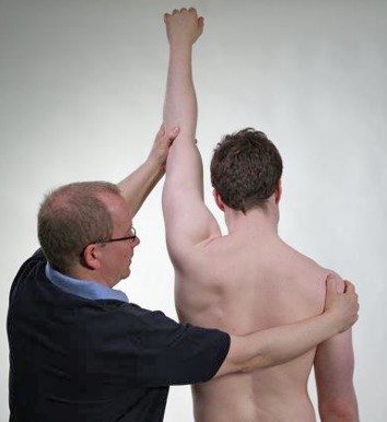
The examiner stabilizes the right shoulder and passively lifts the left arm up toward the ceiling. Note if pain is triggered in the range of motion. Pay particular attention to pain at the end of the range of motion.
Abduction of the shoulder joint with fixed scapula
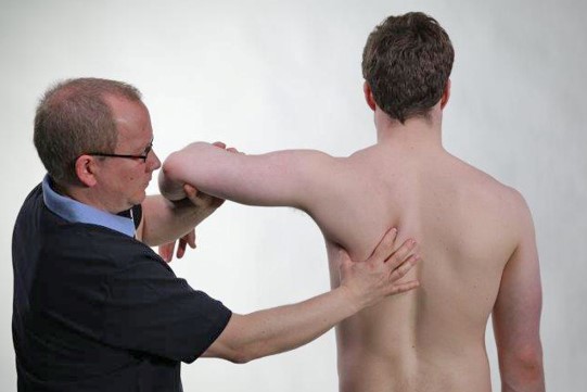
The examiner fixes the tip of the scapula and passively puts the shoulder into abduction. The examiner notes when the scapula begins to move. Normally, the scapula movement starts with about 90° passive abduction.
Humeroscapular movement
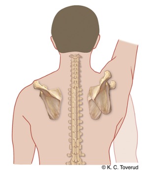
The humeroscapular movement is a total of 180°. The movement is composed of the shoulder joint abdicating a total of 120°, while the scapula rotates 60° on the thorax. This rotational motion of 60° is a sum of rotation and elevation in the sternoclavicular joint of 30° plus rotation in the AC joint of 30°.
Adhesive capsulitis, "Frozen shoulder"
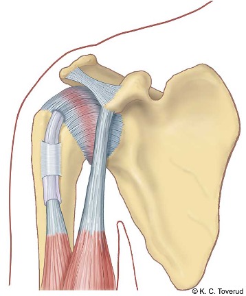
Adhesive capsulitis, also called frozen shoulder, is a painful condition. The condition is caused by an inflammation of the joint capsule, particularly pronounced at the front of the shoulder joint.
Clinical findings during examination in the pain phase (lasting from a few weeks to nine months) may include reduced passive abduction and early movement of the scapula during passive abduction, reduced passive external rotation, and severe pain at the end points of the range of motion (spastic end-feel). In the stiffness phase (from 9 to 15 months), loss of motion dominates, but decreasing pain is found during clinical examination.
External rotation 90°
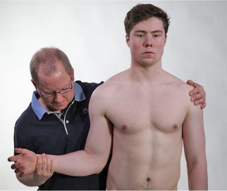
The examiner stabilizes the patient's left shoulder to prevent rotational movement in the body. The examiner moves the right shoulder into maximum external rotation. The examiner pays particular attention to the range of motion and whether there is painful end-feel.
Internal rotation 90°

The examiner stabilizes the patient's back to prevent rotational movement and moves the patient's left shoulder into maximum internal rotation. This position allows checking whether there is painful end-feel by gently lifting the arm away from the back.
Subacromial bursitis
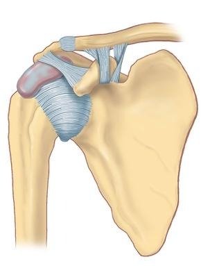
The clinical condition subacromial bursitis is diagnosed when there is a painful arc (painful arc). Pain arc is often seen in combination with normal passive range of motion in the shoulder joint, but with pain at the end point of the range of motion in one or more directions of movement. A constant finding is pain at the end point of maximal passive elevation.
Horizontal shoulder adduction
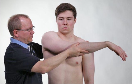
Examination of the acromioclavicular joint (AC joint) is performed by stabilizing the left shoulder and moving the shoulder from 90° flexion toward maximum adduction in the horizontal plane. The examiner records whether the test triggers pain over the AC joint.
Park HB et al. Diagnostic accuracy of clinical tests for different degrees of subacromial impingement syndrome. J Bone Joint Surg Am. 2005;87:1446-1455
Horizontal adduction with compression of the acromioclavicular joint

In case of suspected arthritis or osteoarthritis in the joint, the examination can be enhanced by applying a little extra pressure at the end of the range of motion, so that the joint is compressed. If there is osteoarthritis or arthritis in the joint, this extra compression will often trigger pain (Kibler and Sciascia, 2022).
Nonspecific test, can also be positive in
- Subacromial bursitis
- Rotator cuff rupture
Isometric movements
- Assess if there is muscle weakness and pain.
- Consider if there is a rotator cuff tendinopathy and/or rotator cuff rupture.
Shoulder, innervation
Palpation
- Assess if there is localized pain in the bursa, muscles, or joint.
- Consider about whether there is bursitis in the subdeltoid bursa, a trigger point in the m. infraspinatus, AC joint osteoarthritis, or AC joint dislocation.
m. deltoideus and underlying subdeltoid bursa
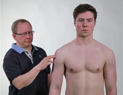
The examiner palpates over m. deltoideus to determine if it triggers pain in the underlying subdeltoid bursa.

m. deltoideus
Subdeltoid bursitis
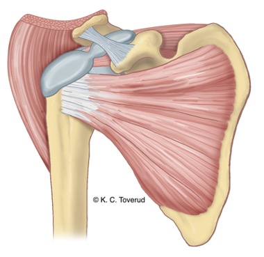
The subdeltoid bursa lies under m. deltoideus.
- The condition starts suddenly. Often unknown cause.
- Pain increase is seen during the first three days, then gradually improves over three to six weeks.
- The patient is unable to move the shoulder due to pain and keeps the elbow close to the body.
- It is not possible to perform a full functional examination, but the most typical findings are markedly limited passive abduction and strong indirect tenderness in the subdeltoid bursa when palpating the deltoid muscle.
m. infraspinatus
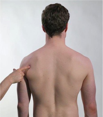
On palpation, pressure is applied to the muscle in the infraspinous fossa. Note whether there is local tenderness in the muscle, and whether a tender pressure point causes referred pain down the upper arm. The latter is a common finding with tension and sore muscle knots in the m. infraspinatus.

m. infraspinatus
Symptoms of fibromyalgia and shoulder myalgia
| Symptom/Finding | Fibromyalgia | Shoulder myalgia |
| Pain location | Often several regions at the same time | Localized, most often in the trapezius and infraspinatus muscles |
| Pain radiation | No, but migration of pains to different parts of the body | No, usually localized |
| Trigger points | Usually not | Usually not, but radiation to the head and arm is common |
| Muscle tenderness | Yes, but often in combination with abdominal pain and headache | Yes, in muscles |
Acromioclavicular joint
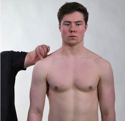
Palpate the joint space of the acromioclavicular joint. The easiest way to find the joint space is to palpate along the anterior edge of the acromion until you reach the joint space approximately 1 cm from the lateral anterior edge of the acromion. Note if there is pain on palpation, and if there is a side difference.
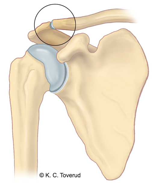
Acromioclavicular joint
Acromioclavicular joint injury
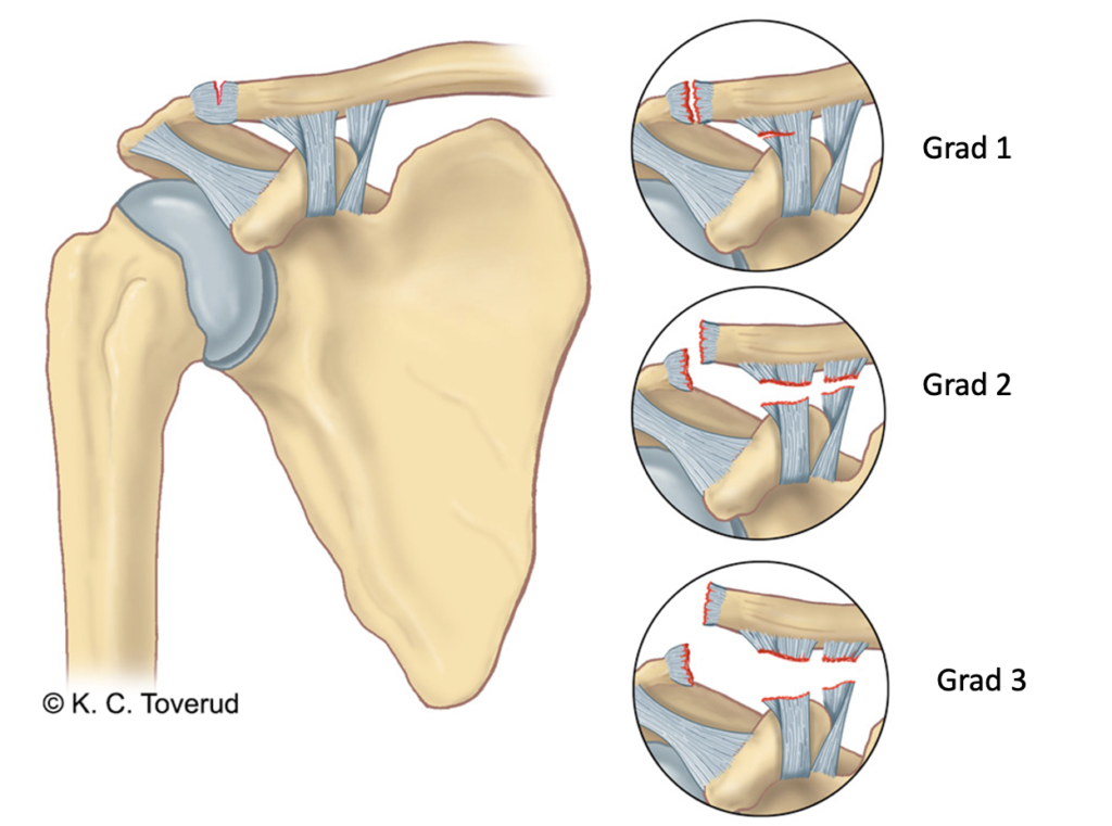
Injury to the acromioclavicular joint usually occurs after a fall trauma. The trauma can cause a dislocation in the joint. The degree of dislocation determines what you find during the clinical examination.
- Grade 1 is damage to the joint capsule and a tear in one ligament. Causes tenderness on palpation in the joint, often with visible joint swelling, but without a clear dislocation.
- Grade 2 and 3 are a combination of damage to the joint capsule and total rupture of the ligaments, resulting in varying degrees of dislocation. Causes tenderness on palpation in the joint and distinctly altered contour in the joint as a sign of dislocation.
Assessment of clinical presentation based on functional examination
You get important information from active abduction of the shoulder. If the patient reports pain during abduction but has a normal range of motion (180°), you must determine whether there is a painful arc (60-120°) or if there is pain at the end point of active abduction. If the patient has a painful arc, you should consider that there may be a subacromial impingement. The structures that can cause impingement are an inflamed subacromial bursa (usually severe pain between 60-120°), inflamed tendon attachment for the supraspinatus and/or infraspinatus (the tendon attachment on the greater tubercle is pinched against the lateral edge of the acromion), or inflamed tendon attachment for the subscapularis (the tendon attachment on the lesser tubercle is pinched against lateral edge of the acromion). Narrowing under the coracoacromial ligament and the lower edge of a worn AC joint with osteophytes are also important causes of impingement.
If it is the subacromial bursa that is the cause of the impingement, in addition to the pain arc, you will often find pain at the end range of motion during passive movement, especially during passive abduction but also during passive external rotation and/or internal rotation.
If it is an inflamed tendon attachment that is causing impingement, you will typically experience a pain arc plus pain during isometric testing of the affected muscle, pain during isometric abduction (supraspinatus), isometric external rotation (infraspinatus), and isometric internal rotation (subscapularis). Special tests for impingement (Neer's test, Hawkins test, Empty can test) are mostly used in orthopedic specialist outpatient clinics.
If you, during active movement examination, find that the patient cannot perform abduction and experiences severe pain already at 20-30° of abduction, you must check this finding further with a passive movement examination. If you suspect that there may be capsulitis, you will find clearly reduced passive abduction with severe pain at the endpoint of the range of motion. During this examination, you must stabilize the scapula to see if early scapular movement is occurring, and compare with the healthy side. In addition to reduced abduction, with capsulitis you will find significantly reduced external rotation with severe pain at the endpoint of the range of motion (the capsular pattern is most reduced external rotation, less abduction, least internal rotation).
If there is information about trauma in the medical history (old trauma or recent trauma), you must consider whether a rotator cuff rupture may be present. In that case, the isometric tests provide the most information, and you must record if there is pain and reduced strength (which may indicate a partial rupture) or only reduced strength (which may indicate a total rupture). Supraspinatus rupture causes reduced strength for abduction in the first 20°, infraspinatus rupture causes reduced strength for external rotation, and subscapularis rupture causes reduced strength for internal rotation.
If the history includes information about shoulder trauma, and possibly previous shoulder dislocation, an apprehension test can be performed in patients who report problems in the form of instability. Some patients without a history of trauma may also have increased joint mobility, which can be found when examining the passive range of motion; a positive sulcus sign may indicate increased joint mobility. Most patients with increased joint mobility have no complaints.
Many patients present with shoulder pain located over the AC joint. Usually, a standard clinical examination of the shoulder when AC joint osteoarthritis is present will be negative. With a negative shoulder examination or when an examination does not reveal a clear pattern suggesting bursitis, tendinopathy, or capsulitis, you must consider AC joint osteoarthritis. You then perform an AC joint test in the form of passive horizontal adduction and note whether it reproduces the patient's pain. You can also perform palpation of the AC joint and compare it with the healthy side.
Special tests
Final competence

- Perform relevant clinical examination in a skilled and gentle manner
- Interpret clinical findings and be able to establish tentative diagnoses
- Suggest referral and treatment
Last updated
31.08.2021