
Inspection
- Assess Gait pattern and obtain an image of the knee joint's dynamics (limping gait, joint range of motion). Look for whether the contours of the knee are symmetrical, especially the tibial tuberosity with regard to Osgood-Schlatter disease. Look for atrophy of the m. vastus medialis; this muscle is weakened in patellofemoral pain syndrome. Assess the leg alignment (valgus/varus position in the knee joint) and evaluate whether the Q-angle (the angle between the m. rectus femoris and the patellar tendon) is within the normal range. Marked varus/valgus position in the knee joint during the stance phase can indicate ligamentous instability or osteoarthritis. Normal Q-angle is 13° in men and 18° in women. Increased Q-angle causes genu valgum, which results in increased lateral pull on the patella and leads to increased risk of chondromalacia patella and patellar dislocation.
- Consider the position of the knee joint (valgus/varus, Q‑angle, patella position, hyperextension) and whether the patient limps. Assess the knee joint's mobility and stability, muscle atrophy, effusion, and perform palpation.
From the front

Assess: Leg alignment, Q‑angle, patella position.
From the back
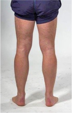
Assess: Axis deviation, hamstring symmetry.
From the side

Assess: Hyperextension.
Medially

Consider: Quadriceps atrophy.
Q-angle

Consider: Q-angle normally 13° in men and 18° in women. Corresponds with a normal physiological valgus in the knee joint.
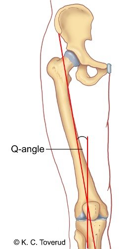
The Q-angle is between the rectus femoris muscle and the patellar tendon
- With genu valgum (knock-knee) the Q-angle increases
- Q-angle outside the normal range is associated with the development of chondromalacia patella and the risk of patellar dislocation
Active movements
- Assess the patient's ability and willingness to perform active mobility in the knee joint. Interpret the findings in light of the patient's age, activity level, and findings from inspection.
- Consider range of motion, pain, and crepitus. Compare findings from the right and left knee joints to identify functional differences. Assess mobility with the patient in a supine position on the examination bench and ensure that the hip and pelvis are stabilized and that the movement is isolated to the knee joint.
Flexion
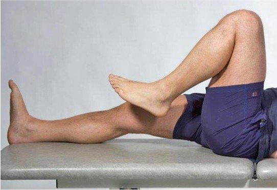
The examiner asks the patient to actively bend the knee joint from a straight position and records range of motion and pain.
Extension
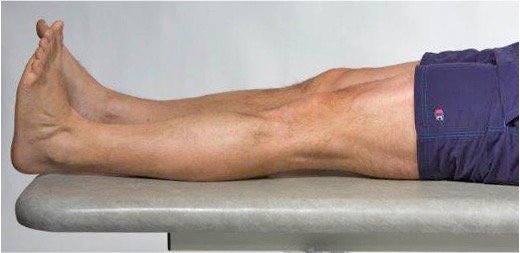
The examiner asks the patient to actively stretch the knee joint from a straight position and records the range of motion.
Passive movements
- Assess Range of motion and indicate degrees, pain, end-feel, and any crepitus. Stabilize the femur and hip so that the movement is isolated to the knee joint. Test both flexion, extension, and rotation. Rotational movements are sensitive to meniscus and cruciate ligament involvement. Increased rotation may indicate cruciate ligament injury, and reduced rotation is seen in osteoarthritis.
- Consider range of motion and indicate degrees, end-feel, pain, and lateral stability. Interpret the findings in light of inspection and active movement. Knee osteoarthritis results in reduced passive mobility in a capsular pattern with more reduced flexion than extension, often combined with a hard end-feel when the passive ranges of motion are examined. The collateral ligaments of the knee are first tested with the knee extended and then with the knee in 30° flexed position.
Flexion 115-160°

The examiner moves the patient's knee from full extension to full flexion. Range of motion, pain, and end-feel at maximum flexion are recorded.
Extension 0-10°
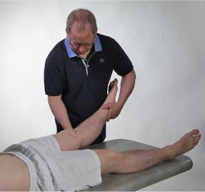
The examiner moves the patient's knee joint from full extension and examines whether it is possible to move the knee joint into hyperextension. Degree of hyperextension, pain, and end-feel in hyperextension are recorded.
Degrees of passive knee mobility
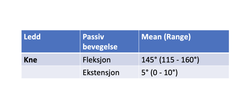
of the Hip, Knee and Ankle Joints in Male Subjects, 30–40 Years of Age, Acta Orthopaedica Scandinavica, 53:2, 205-208
Knee joint osteoarthritis
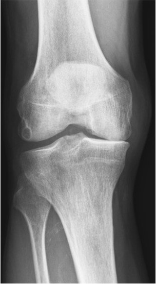
X-ray of medial knee osteoarthritis
The osteoarthritis causes changes in the cartilage and narrowing of the joint space.
Internal rotation 10-15°
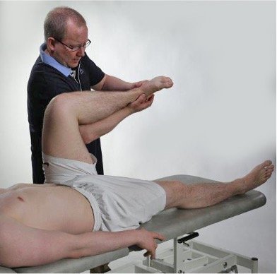
The examiner passively flexes the knee joint to 90° and rotates the knee joint inward. Degree of internal rotation, pain, and end-feel are recorded.
External rotation 20-30°
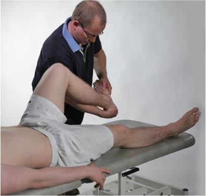
The examiner passively flexes the knee joint to 90° and rotates the knee joint outward. Degree of external rotation, pain, and end-feel are recorded.
Test for pain in the lateral collateral ligament.
With the knee extended, the joint is side-stabilized by the ligaments of the posterior capsule, the posterior and anterior cruciate ligaments, and the popliteus muscle.
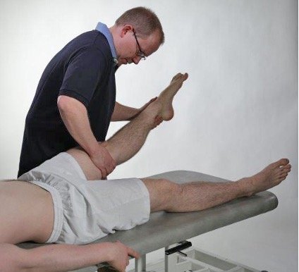
Varus stress is performed. The examiner records whether the test causes pain laterally.
Test for instability in the lateral collateral ligament.
When the knee joint is flexed to 30°, the collateral ligaments become the primary stabilizers.

Varus stress tests the lateral collateral ligament. The examiner records whether the tests indicate lateral instability.
Test for pain in the medial collateral ligament.
With the knee extended, the joint is side-stabilized by the ligaments of the posterior capsule, the posterior and anterior cruciate ligaments, and the popliteus muscle.
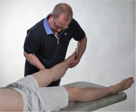
Valgus stress is performed. The examiner records whether the test causes medial pain.
Test for instability in the medial collateral ligament.
When the knee joint is flexed to 30°, the collateral ligaments become the primary stabilizers.
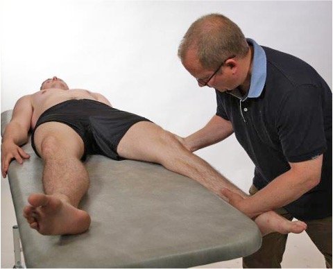
Valgus stress tests the medial collateral ligament. The examiner records whether the tests indicate medial instability.
Anatomy, ligaments, and muscle attachments
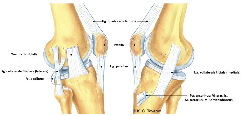
Medial stabilizing structures
- Primary stabilizer
- Medial collateral ligament
- Secondary stabilizers
- Posterior medial capsule
- Semimembranosus muscle
- Anterior and posterior cruciate ligaments
- Pes anserinus (sartorius muscle, gracilis muscle, semitendinosus muscle)
- Medial head of gastrocnemius muscle
- Compression of lateral meniscus
Stabilizing structures laterally
- Primary stabilizer
- Lateral collateral ligament
- Secondary stabilizers
- Posterior lateral capsule
- Popliteus muscle
- Anterior and posterior cruciate ligaments
- Iliotibial tract
- Lateral head of gastrocnemius muscle
- Compression of medial meniscus
Anatomy, ligament injury causes lateral instability

An injury to the collateral ligaments can be isolated or occur in combination with meniscus and cruciate ligament injury. Injury of the medial collateral ligament is the most common knee injury following a twisting trauma and often occurs as an isolated injury. An injury to the lateral collateral ligament is usually associated with simultaneous injury to the meniscus and cruciate ligament
Isometric movements
- Assess to perform an isometric test with the knee joint in 90° flexion for flexion and rotation and in slight flexion for extension. This is so that the muscles work in optimal positions. Stabilize the femur and hip so that the force comes from the correct muscle group. Knee flexion tests the hamstrings, extension tests the quadriceps, internal rotation tests the semitendinosus, semimembranosus, sartorius, gracilis, and popliteus, and external rotation tests the biceps femoris.
- Consider about whether there are clinical signs of hamstring injury (pain at the back of the thigh), quadriceps rupture/tendinopathy (pain at the front of the thigh), pes anserinus tendinopathy (pain medial upper part of the tibia), popliteus tendinopathy (pain at the lateral femoral condyle) or biceps femoris tendinopathy (pain at the lateral tibial condyle, fibular head).
Hamstring injury
- Strain injury, tearing of muscle fibers.
- Usually the tearing occurs at the transition between muscle and tendon.
- Bleeding, which in many cases can be significant at the back of the thigh.
- In some cases, the strain injury causes the attachment to the bone on the pelvic rim, the ischial tuberosity, to be torn off
- The muscle detaches from the pelvis (proximal hamstring avulsion injury)
- Can occur with severe bending at the hip joint while the knee is extended
- Often overlooked early in the course
- Biceps tendinitis and avulsion injury of the attachment on the fibula
- Result of overuse or injury with detachment of the attachment on the fibula (X-ray)
Engebretsen L. I: Sports injuries (Bahr R, Mæhlum S, eds.). Hamstring injury – strain injury on the back of the thigh. Oslo: Gazettebok, 2002.
Pes anserinus tendinopathy and bursitis

- The tendon formed by the three muscles m. gracilis, m. sartorius, and m. semitendinosus
- The tendons of these three muscles unite in a common tendon on the medial tibia, internally rotating the knee joint
- Pes anserinus tendinopathy and bursitis denote an inflammation in the tendon or underlying bursa
- The symptoms are aching pain on the inside of the calf just below the knee. The pain worsens when walking in stairs or when squatting, or after running and jumping
Palpation
- Assess perform palpation systematically. Start suprapatellar and then continue to → patella → patellar tendon → joint spaces → collateral ligaments → posterior structures. Use the back of the hand to assess warmth, as it is more sensitive to temperature differences. Identify localized tenderness, which is often more diagnostic than pain on movement. Compare palpation findings with previous findings from inspection, active/passive movement, and isometric tests. Use palpation as the last part of the examination, unless there is obvious swelling, warmth, or trauma that requires early assessment. Interpret the findings in light of age, activity level, and injury history.
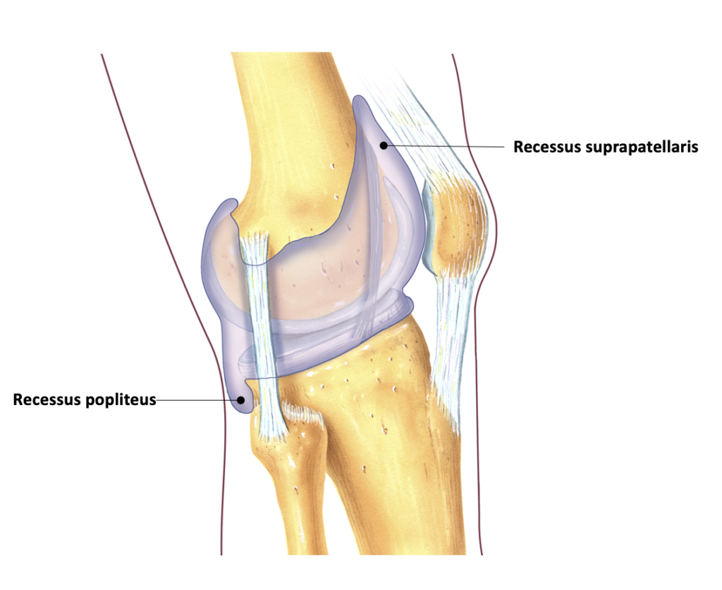
- Consider pain and/or possible rupture in the quadriceps tendon, tenderness over the patella (prepatellar bursitis), warmth (arthritis/bursitis), patella dip test (positive test with >20 ml intra-articular fluid accumulation), presence of a Baker's cyst (secondary to osteoarthritis/arthritis/meniscus rupture), collateral ligaments (tenderness at attachment on femur and/or tibia), jumper's knee (tenderness at patella apex), runner's knee (tenderness at iliotibial tract) or chondromalacia patella/patellofemoral pain syndrome (Patellar Grind Test, Clarke’s test).
Palpation of the patella simultaneously with squatting. Crepitation. Pain.
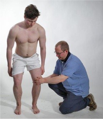
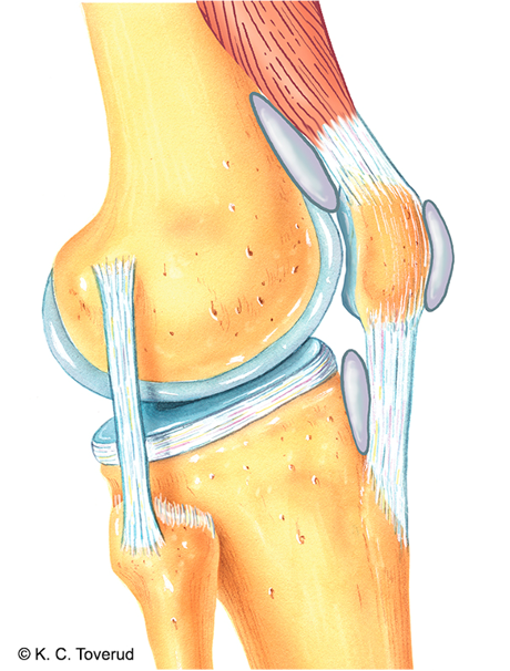
Pain from
- Flaked joint cartilage in the femoropatellar joint
- Prepatellar bursa, suprapatellar, infrapatellar
- Quadriceps tendon
- Patellar tendon
The examiner can palpate over the patella while the patient performs an active squat. The examination provides information about crepitus in the knee joint and whether slight pressure on the patella during the squat triggers pain. Crepitus and pain may indicate prepatellar pain due to prepatellar bursitis, or there may be femoropatellar osteoarthritis or frayed patellar cartilage.
Heat
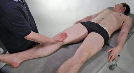
The examiner uses the back of the hand to check for increased local warmth over the knee joint, especially if there is suspicion of arthritis or bursitis in the knee joint.
Patella dip test, positive when > 20 ml intra-articular fluid

Fluid is collected in the knee joint from the suprapatellar recess with one hand and from the infrapatellar structures with the other hand. Once this is done, the index finger is used to press the patella down against the femur and it is noted whether the patella rises again – "dips" – when the pressure against the patella is released. A rule of thumb is that the patella dip test is positive if there is more than 20 ml of intra-articular fluid in the knee joint.
Anatomy, suprapatellar recess and popliteus
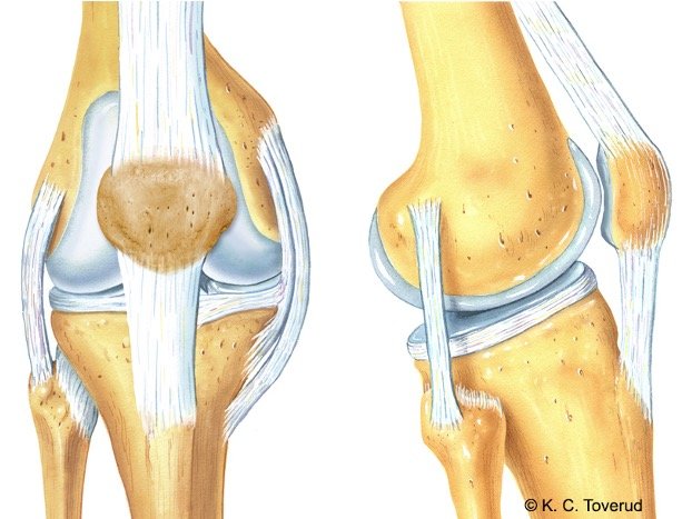
Medial collateral ligament
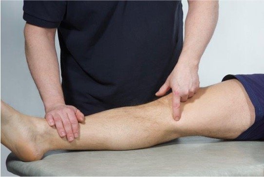
In case of suspected medial collateral ligament injury (ligamentum collaterale mediale), one palpates for tenderness in the joint space with the knee extended and with the knee joint at 90° flexion. The ligament runs from the femur down to the tibia and is best palpated from the midline of the joint and further dorsally. If there is significant pain upon palpation at the ligament's attachment on the femur and/or on the tibia when the examination is performed with the knee extended, the suspicion of ligament injury is increased (Braaten et al., 2022). If the pain is mainly localized in the joint space itself in both extended and flexed knee positions, a meniscus injury should be suspected.
Lateral collateral ligament
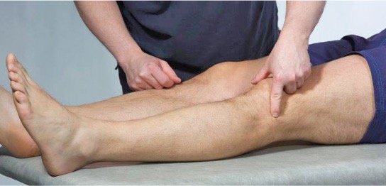
In case of suspected lateral ligament injury (lateral collateral ligament), one palpates for tenderness in the joint space with the knee extended and with the knee joint in 90° flexion. The ligament runs from the femur down to the head of the fibula and is best palpated from the midline of the joint and further dorsally. If there is significant pain when palpating the ligament's attachment on the femur and/or on the fibula when the examination is done with the knee extended, the suspicion of a ligament injury is strengthened. If most of the pain is located in the joint space itself both in the extended and flexed knee, one should suspect that a meniscus injury may be present.
Anatomy, collateral ligament
Medial
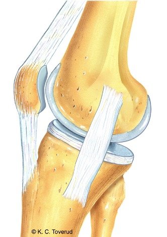
Lateral
Patellar tendon
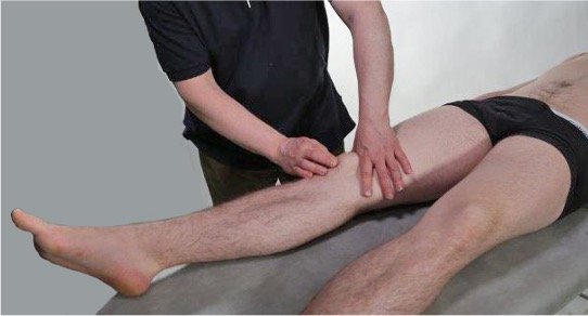
The examiner presses the patella down at the upper edge, so that the apex of the patella is lifted and the attachment of the patellar tendon becomes accessible for palpation. Pain during palpation strengthens the suspicion that the condition is present.
Iliotibial tract
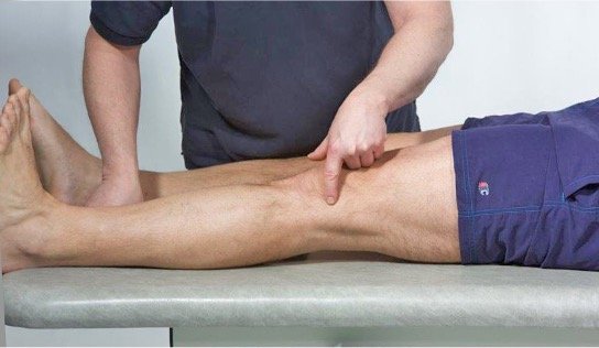
With clinical suspicion of jumper's knee with moderate to severe pain, the patient can be examined by palpation of the iliotibial tract tendon and the underlying bursa. On examination, local tenderness and crepitation over the bursa are often found upon palpation.
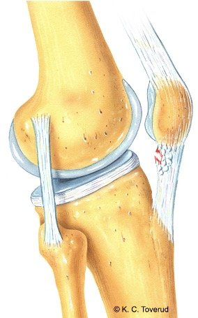
Patellar tendon
The patellar tendon, location jumper's knee “Jumper’s knee”
Jumper's knee is part of the clinical picture we call patellofemoral pain syndrome. The condition is caused by small degenerative changes in the patellar tendon, especially at the attachment on the apex of the patella. Jumper's knee is a common condition among those who engage in sports, particularly volleyball and football.
Iliotibial tract
Iliotibial tract with bursa, location runner's knee “Runner’s knee”
Runner’s knee is part of the clinical picture of patellofemoral pain syndrome. The condition is caused by inflammation in the iliotibial tract tendon or in the underlying bursa at the lateral femoral condyle where the tendon structure passes before attaching to the lateral tibial condyle. The condition is characterized by activity-related pain, especially during running. It is treated with stretches with a physiotherapist. Steroid injection in the bursa can be an option for long-term complaints.
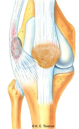
Baker’s cyst
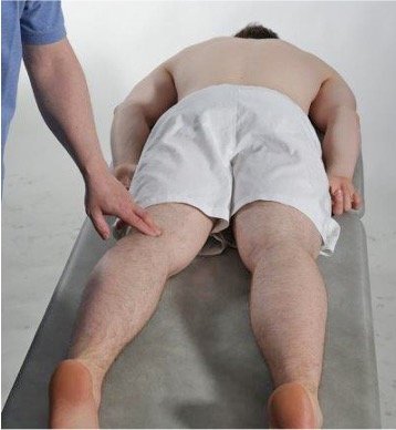
Intra-articular pathology such as osteoarthritis, arthritis, or degenerative meniscus rupture can cause increased intra-articular fluid accumulation. Over time, this will lead to increased intra-articular pressure. The pressure increase can result in the formation of a so-called Baker's cyst in the popliteal fossa. The cyst is usually asymptomatic, but if it ruptures, the patient may experience acute increased pain in the popliteal fossa with referred pain and possibly swelling and redness in the calf. The condition can be confused with deep vein thrombosis.
Palpation of the popliteal fossa
Baker’s cyst
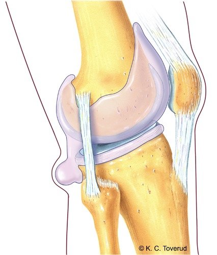
Baker's cyst, location

Ruptured Baker's cyst
- -Occurs in patients with known knee joint problems
- Rheumatic disease
- Osteoarthritis
- Degenerative meniscus disorder
- Acute pain, swelling in the calf, and ankle edema
- Differential diagnosis is deep vein thrombosis (DVT)
Assessment of clinical presentation based on functional examination
Many conditions in the knee joint are triggered by trauma. It is therefore very important that in the medical history, one takes into account old or possibly recently occurred traumas. In the older population, knee joint osteoarthritis is the most common condition. The clinical picture is composed of pain during activity and often joint swelling. On clinical examination, there is often a capsular movement pattern on passive tests (more reduced flexion than extension). Hydrops is tested with the patella dip test.
With a non-capsular movement pattern, one must consider meniscus injury after trauma or meniscus degeneration as we often see in an arthritic joint. Here, passive extension is often reduced and painful at the end of the range of motion because the meniscus can be pinched in the joint space during extension. If there is suspicion of a meniscus injury, meniscus tests must be performed, usually the McMurray test. A non-capsular movement pattern can also be found with a loose body in the knee joint or if there is a ligament injury (most often the medial collateral ligament) or a prepatellar bursitis.
If there is a normal range of motion and negative isometric tests, one must consider that there may be patellar chondromalacia and perform a Grinding test to determine if it is the cause of the pain.
If there is a normal range of motion and pain during isometric extension in the knee joint, one must consider tendinopathy and/or partial ruptures in the quadriceps muscles around the knee joint; if there is pain during isometric flexion, one must consider injury to the hamstring muscles.
If the patient reports trauma to the knee joint or a known cruciate ligament injury, it is important to examine stability. The anterior cruciate ligament is tested with the "Drawer Test" and the medial and lateral collateral ligaments with valgus and varus pressure on a slightly flexed knee joint.
Special tests
Final competence

- Perform relevant clinical examination in a skilled and gentle manner
- Interpret clinical findings and be able to establish tentative diagnoses
- Suggest referral and treatment
Last updated
31.08.2021