
Inspection
- Assess about whether there are external signs of injury, deformity, or deviation in the elbow angle. Swelling and discoloration may be due to acute injury, bleeding, or inflammation. Intra-articular swelling points to joint pathology, diffuse swelling to soft tissue injury. Malalignment indicates fracture or dislocation.
- Consider about whether there is normal or reduced extension position in the elbow joint. Lack of extension may be due to intra-articular foreign body, capsular contracture, or previous injury. Normal valgus angle in the elbow joint is 13–16° for women, 11–14° for men. Cubitus valgus (>15°) → typically after lateral epicondyle fracture and cubitus varus (<10°) → typically after supracondylar humerus fracture.
Extension
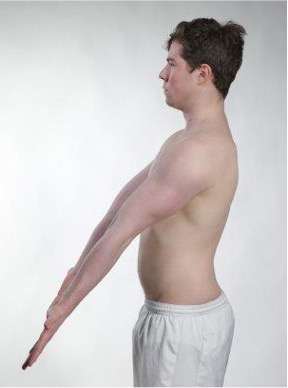
Stand in front of the patient and assess the elbow in full extension. Look for swelling, redness, discoloration, deformity, and symmetry.
Valgus angle
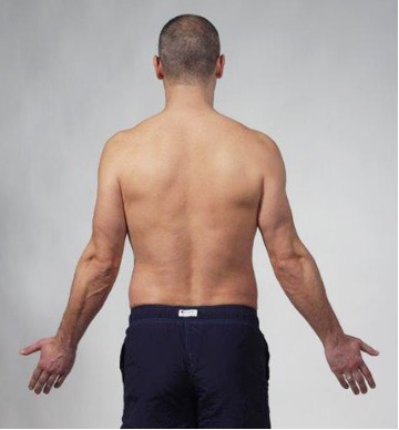
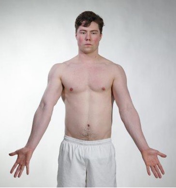
Measure or estimate the valgus angle and assess whether it is within the normal range. Note if there is cubitus valgus or cubitus varus, and evaluate whether this aligns with the patient's medical history.
Active movements
- Assess the patient's ability and willingness to move the elbow joint. If the patient reports pain, assess where in the range of motion the pain occurs (start, middle, or at the end point of the range of motion).
- Consider about the range of motion in the elbow joint is normal, reduced, or asymmetric. Try to distinguish between pain, mechanical block, and reluctance. Reduced flexion is seen in capsular contracture, osteoarthritis, or pain. Reduced extension is often the first sign of intra-articular pathology. Limited pronation is seen in fracture, instability, or muscular tightness. Reduced supination is seen in dislocation, fracture, or nerve injury. If there is pain, consider whether the pain may be due to tendinopathy (pain with active use), arthritis or bursitis (joint swelling and pain throughout the range of motion), trauma (malalignment and reduced range of motion), or neurological involvement (unusual movement patterns and weakness without pain).
Flexion

Extension

Pronation
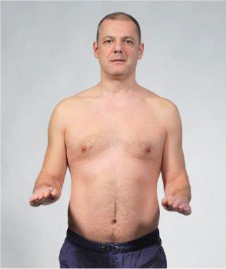
Supination

Passive movements
- Assess whether passive movement in the elbow joint is normal, reduced, or painful and whether this differs from findings in active movement. Assess whether pain occurs at the start, middle, or end point of the movement. This is important to distinguish between capsular pathology, bursitis, arthritis, and mechanical blockage of the range of motion.
- Consider whether the end-feel is normally elastic, hard (normal at full extension, but can also occur early in the range of motion in cases of mechanical blockage, osteophytes, or fracture sequelae) or spastic/painful (capsulitis or arthritis).
Flexion 140-170°
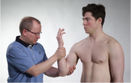
Flexion is tested by holding a firm grip on the patient's forearm and flexing the forearm.
Extension 0-10°
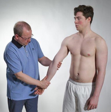
Extension is tested by placing one hand under the elbow joint and moving the forearm into full extension.
Pronation 80°
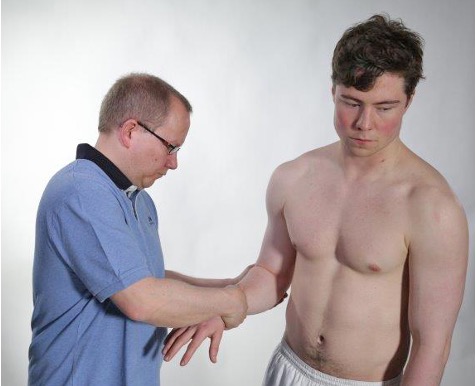
Pronation is tested by holding a firm grip on the forearm, the other hand placed over the wrist, and moving the forearm into full pronation.
Supination 90°
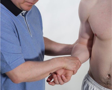
Supination is tested by holding a firm grip on the forearm, the other hand placed over the wrist, and moving the forearm into full supination.
Passive movement tests joints, capsules, cartilage, and ligaments. The examination is crucial for distinguishing between contractile (muscles and tendons) and non-contractile (joint capsule, ligaments, and bursa) pathology. If both passive and active movement are reduced, this points to joint pathology such as capsulitis, arthritis, or mechanical blockage (loose bone or cartilage body).
Osteochondritis dissecans (loose bone or cartilage body)
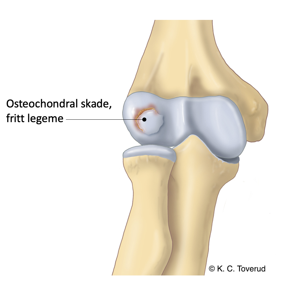
Osteochondritis dissecans typically causes catching, locking, crepitation, and reduced extension. The joint may be warm and swollen. Pain often occurs radially over the capitulum humeri. The clinical finding is confirmed with MR arthrography and CT.
- Osteochondral fragment (avascular necrosis causing subchondral fracture and fragmentation) usually from the capitulum of the humerus.
Valgus stress/stability
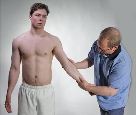
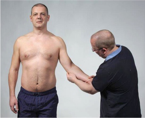
The medial collateral ligament is tested with valgus stress. You place one hand radially over the elbow and the other hand ulnarly over the wrist. Perform a valgus pressure against a straight elbow, and note if it triggers pain or feels unstable.
Varus stress/stability
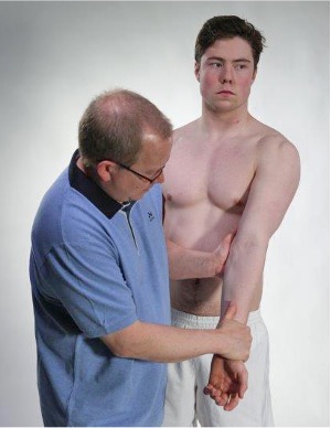
The lateral collateral ligament is tested with varus stress. You place one hand ulnarly over the elbow and the other hand radially over the wrist. Apply a varus pressure with the elbow straight, and note whether it triggers pain or feels unstable.
Valgus and varus stress provide information about the stability of the elbow joint. The examination is important for mapping elbow complaints that may be caused by ligament injuries or instability following trauma and sports injuries.
Isometric movements
- Assess whether an isometric test triggers pain, weakness, or both. Assess whether the pain pattern matches previous findings from active and passive movement. Assess whether there are side differences that may explain loss of function or load-related complaints.
- Consider on clinical findings pointing towards tendinopathy (pain without loss of strength), partial rupture (pain plus reduced strength), total muscle/tendon rupture (reduced strength without pain) or neuropathy (weakness in muscles that share the same spinal nerve or peripheral nerve).
Palpation
- Assess deviations from normal anatomy and localize swelling, heat, redness, crepitus or tenderness using palpation. Systematically palpate the ulnar nerve in the ulnar groove, the olecranon bursa, proximal radioulnar joint, medial and lateral epicondyle.
- Consider about n. ulnaris is tender or irritated in the ulnar sulcus (paresthesia in the 4th-5th finger), whether the olecranon bursa is swollen, warm, red, or painful (bursitis), whether the proximal radioulnar joint causes pain or instability during rotation and compression (injury to the annular ligament of the radius), or whether there is tenderness in the lateral epicondyle (tennis elbow) or medial epicondyle (golfer's elbow).
Ulnar sulcus
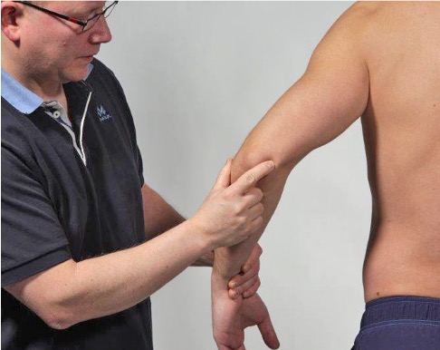
The ulnar nerve lies superficial in the ulnar sulcus and can easily be irritated by pressure or repetitive strain. Assess whether pressure causes paresthesia or pain.
Ulnar nerve entrapment
Olecranon bursa
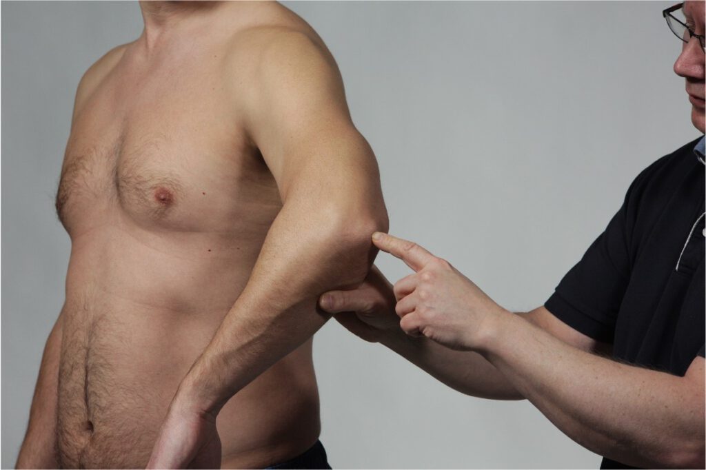
The olecranon bursa is very superficial and therefore prone to trauma, pressure, and infection.
Olecranon bursitis (Student elbow)

Assess the degree of swelling, redness, and warmth. Often tender on palpation. There may be reduced flexion ability in the elbow.
Pulled elbow
Reposition

In pulled elbow, the child holds the elbow in a pronated and extended position. The child refuses to move the forearm into supination.

The repositioning maneuver is to move the forearm simultaneously into supination and flexion.
Move the forearm into supination and flexion
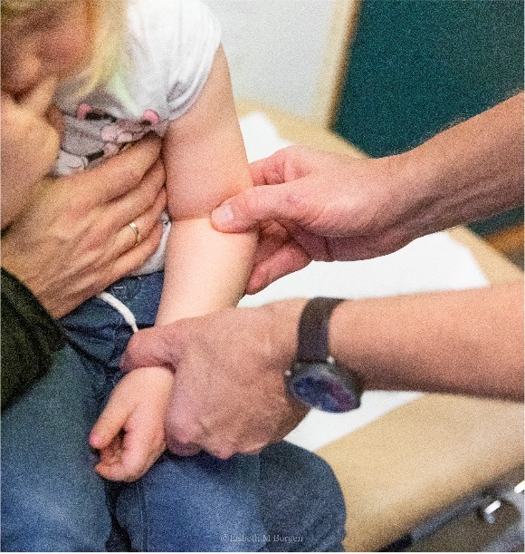

Pulled elbow
Mechanism of injury
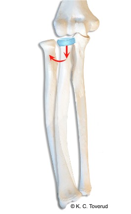
- Pull on the arm that causes the radial head to come out of position from the annular ligament. The elbow remains in extended and pronated position
Proximal radioulnar joint
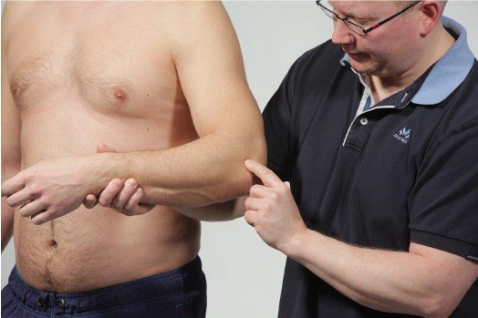
Stability in the proximal radioulnar joint is tested with anterior pressure on the radial head while the elbow
is extended and the forearm rotated. Test for rupture of the annular ligament in children and in
Moteggia fracture (ulna fracture and dislocation of the radial head)
Medial humeral epicondyle
Ulnar epicondylalgia is palpated at the ulnar humeral epicondyle at the attachment for the wrist flexor muscles.
Radial humeral epicondyle

Radial epicondylalgia is palpated at the radial humeral epicondyle at the attachment for the wrist extensor muscles.
Assessment of clinical presentation based on functional examination
In elbow examination, you obtain a lot of information by examining passive mobility. If the range of motion is reduced, you must determine whether the reduced range of motion has a capsular pattern (more reduced flexion than extension); this may indicate arthritis in the elbow joint. If the range of motion is reduced in a non-capsular pattern (for example, clearly reduced extension), you should consider whether there may be a loose body in the elbow joint (a piece of cartilage or bone/cartilage that has separated due to avascular necrosis or trauma).
If there is a normal range of motion in the elbow joint and negative isometric tests of the muscles, you should consider whether there may be olecranon bursitis. There will often be swelling, redness, and warmth over the olecranon bursa with tenderness on palpation; if the inflammation is severe, the patient may report pain during passive flexion.
If during the elbow examination you find that the range of motion is normal but the isometric tests are positive, there may be tennis elbow or golfer's elbow.
In suspected tennis elbow, you will find a positive test with isometric extension of the wrist; if you also find positive isometric radial deviation of the wrist, it may indicate involvement of the tendon attachment on the radial humeral epicondyle for the extensor carpi radialis longus and brevis muscles. If, in addition to pain during wrist extension, you also experience pain with ulnar deviation of the wrist, it may indicate involvement of the tendon attachment for the extensor carpi ulnaris muscle.
Positive isometric flexion in the wrist may indicate golfer's elbow with involvement of the tendon attachment for the flexor musculature at the attachment on the ulnar humeral epicondyle.
Special tests
Final competence

- Perform relevant clinical examination in a skilled and gentle manner
- Interpret clinical findings and be able to establish tentative diagnoses
- Suggest referral and treatment
Last updated
31.08.2021