
Inspection
- Assess the patient in a standing position. Note whether there is a pelvic tilt where the pelvis is rotated forward with a pronounced lumbar lordosis (often associated with tight m. psoas, m. erector spinae, and m. QL), which can cause lower back pain due to increased muscular load, or whether the pelvis is rotated backward with a flattened lumbar lordosis (can be compensation for pain, stiffness, or weakness in the back/hip muscles). Identify the anterior superior iliac spine (ASIS is the front hip point, attachment for m. sartorius, TFL, and inguinal ligament), anterior inferior iliac spine (AIIS is the upper edge of the acetabulum, attachment for m. rectus femoris and m. iliacus), and posterior superior iliac spine (PSIS is the posterior edge of the iliac crest). attachment for m. multifidi and lig. sacroiliaca dorsalia) precisely, as these landmarks form the basis for assessing pelvic position, muscle attachments, and ligament structures.
- Consider whether there are signs of asymmetry, flattening, hyperlordosis, rotation, or misalignment, which may explain pain patterns in the lower back and pelvis. Muscles that maintain lumbar lordosis are the m. multifidus, m. erector spinae, m. quadratus lumborum, and m. psoas major. The iliosacral joints are partly a synovial joint (about 25% of the joint surface), the remaining part of the joint is referred to as a syndesmosis. A syndesmosis is a joint connection, a false joint without a joint cavity, held together by connective tissue and is immobile. The joint is stabilized by strong ligaments, lig. sacroiliaca anterior and posterior. Consider whether the pain primarily originates from ligament structures or from muscles.
Anterior pelvic tilt

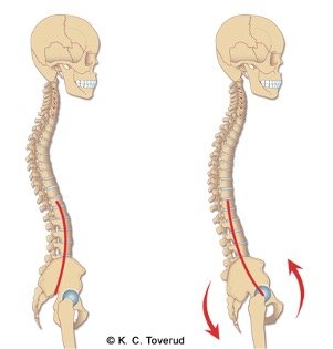
Assess whether there is an anterior pelvic tilt and increased lumbar lordosis. Anterior pelvic tilt is associated with lower back pain. It is assumed that the pain comes from tense muscles working to maintain the posture.
Chun SW, Lim CY, Kim K, Hwang J, Chung SG. The relationships between low back pain and lumbar lordosis: a systematic review and meta-analysis. Spine J. 2017 Aug;17(8):1180-1191. doi: 10.1016/j.spinee.2017.04.034.
Posterior pelvic tilt
- With posterior pelvic tilt, there is
- Postural flattening (bending) of the lower back
- The patient tightens the stomach, buttocks, or hamstrings to create stability in the pelvis
- The flattening of the lower back will result in reduced function of the muscles that maintain the curve in the lower back
- Posterior pelvic tilt can cause the body's center of gravity to move forward
- Hangs backward in the middle back
- Locks the knees
- Forward tilt of head and shoulders
- Hyperextension in the hip joints
ASIS (Anterior Superior Iliac Spine)

- Localized anterior edge of the ilium
- Forms upper edge of the acetabulum
- Attachment for
- m. rectus femoris
- m. iliacus
- Lig. iliofemorale
PSIS (Posterior Superior Iliac Spine)

- Localized posterior edge of the iliac crest
- Attachment for
- Mm. multifidi
- Dorsal sacroiliac ligaments
Active movements
- Assess Flexion can trigger localized pain in the iliosacral joints or referred pain to the iliac fossa, gluteal area, groin, or along the lateral side and back of the thigh down to the knee joint area. If there is suspicion of rheumatic disease (spondyloarthropathy), the Schober test should be performed. The test quantifies the degree of reduced segmental lumbar flexion movement when inflammatory spinal disease is suspected.
- Consider about the mobility of the pelvic joint by placing one finger on each PSIS to record whether the movement is symmetrical and whether it triggers pain. The assessment is uncertain if performed as a single test; a positive finding must be considered together with other pelvic joint tests.
Flexion
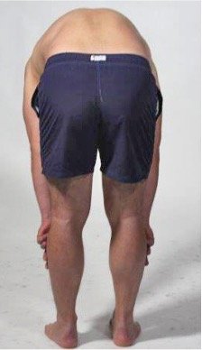
Have the patient bend forward. Note whether flexion triggers localized pain in the sacroiliac joints or referred pain.
Passive movements
- Assess about axial pressure over the sacrum triggers pain. Place the patient in a prone position on a stable bench so that the ilia are fixed and the sacrum can move relative to the pelvis. The pressure creates an anterior shearing force on both sacroiliac joints. Be aware that the test can provoke both the sacroiliac joint, ligaments, and deep gluteal structures, and a positive test must therefore be considered together with other findings.
- Consider whether the test causes unilateral pain over one iliosacral joint (typically with wear and tear) or bilateral pain (can occur in inflammation-like rheumatic disease). Pain that is not recognizable to the patient speaks against the iliosacral joint as the primary source of pain. A single positive test has limited value, but a negative test in combination with other negative tests speaks against iliosacral joints as a source of pain. Lumbar disc pathology causes pain more centrally, often with radiation.
- Facet joint osteoarthritis causes pain during extension, not sacral thrust. Gluteal myalgia or piriformis syndrome causes more lateral pain. The iliolumbar ligament causes more cranial pain over the iliac crita.
Pressure against the sacrum (sacral trust test)
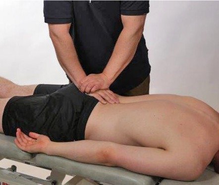
Press against the sacrum with the patient in the prone position. An axial downward pressure is applied. Note whether the patient reports unilateral or bilateral pain over the sacroiliac joints.
Palpation
- Assess to perform systematic and precise palpation to identify local tenderness, swelling, asymmetry, muscle tension, or pain radiating from tender structures in joints, ligaments, or muscles. It is important to know which muscles and ligaments attach at each landmark so that you can link palpation findings to likely clinical causes.
- Consider swelling or increased tenderness over the pubic symphysis that may indicate inflammation or overuse. If there is pain with adduction or abdominal contraction, this points to myotendinous causes (m. rectus abdominis, m. adductor longus). Then evaluate findings on palpation over the pubic tubercle; pain during simultaneous active or isometric adduction will strengthen the suspicion of adductor pathology. Local pain over the ischial tuberosity may be due to hamstring tendinopathy (m. semimembranosus, m. semitendinosus, m. biceps femoris) or ischiogluteal bursitis. Pain on palpation combined with active or isometric knee flexion strengthens the suspicion of hamstring pathology.
Pubic symphysis

- Non-synovial joint with fibrocartilage
- The stability of the joint is ensured by ligaments
- Pubic symphysis attachment for
- m. rectus abdominis
Pubic tubercle
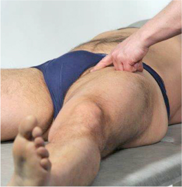
- Located medially on the superior ramus of the pubic bone. Important structure in adductor-related groin pain.
- Attachment for
- m. adductor longus
- m. gracilis
- Lig. inguinale
Ischial tuberosity
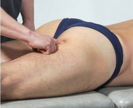
- Located posteriorly on the superior ramus of the ischium. Important structure to consider in hamstring avulsion and in tendinopathy.
- Attachment for
- m. adductor magnus
- m. semimembranosus
- m. semitendinosus
- m. biceps femoris, long head
- Lig. sacrotuberale
Assessment of clinical presentation based on functional examination
Functional examination of the pelvis with the sacroiliac joint is performed as part of a screening examination when there are low back pains and pain in the hip area. The goal of the examination is to map whether the patient's pain originates from the sacroiliac joint and whether arthritis in the joint, as seen in inflammatory rheumatic disease, can be suspected. The clinical examination must be structured so that it is determined whether the pain may come from the lumbar spine or the hip joint before concluding that sacroiliac joint arthritis may be present. It is important to focus on the sacroiliac joint in younger people who present with low back pain. In older people, it is often a more complex clinical image where contributions to pain in this region can partly come from degenerative changes in the lumbar spine, early hip joint osteoarthritis, and degenerative changes in the sacroiliac joint. The sacroiliac joint tests are designed to diagnose arthritis but can be false positive with degenerative joint changes.
Special tests
Last updated
31.08.2021