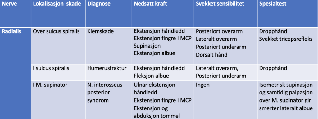
N. interosseus posterior syndrome, Anatomy
N. interosseus posterior is compressed in 60-70% of cases at Frohse’s arcade (upper edge of M. supinator). Less commonly in the radial tunnel, which extends from the upper edge of M. supinator and 5 cm distally
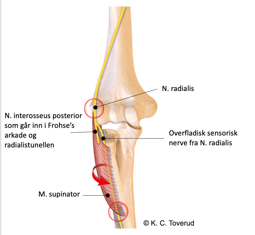
Frohse’s canal starts after the branch to the M. supinator
N. interosseus posterior syndrome, Clinic
N. interosseus posterior syndrome, Clinic
- Caused by trauma, diabetic mononeuropathy, pressure from lipoma, ganglion, or hypertrophic synovitis in RA.
- Pain in the lateral elbow region radiating distally; the nerve has no sensory function, so no sensory loss is detected in the skin. Pain can be provoked by compression 5 cm distal to the lateral humeral epicondyle.
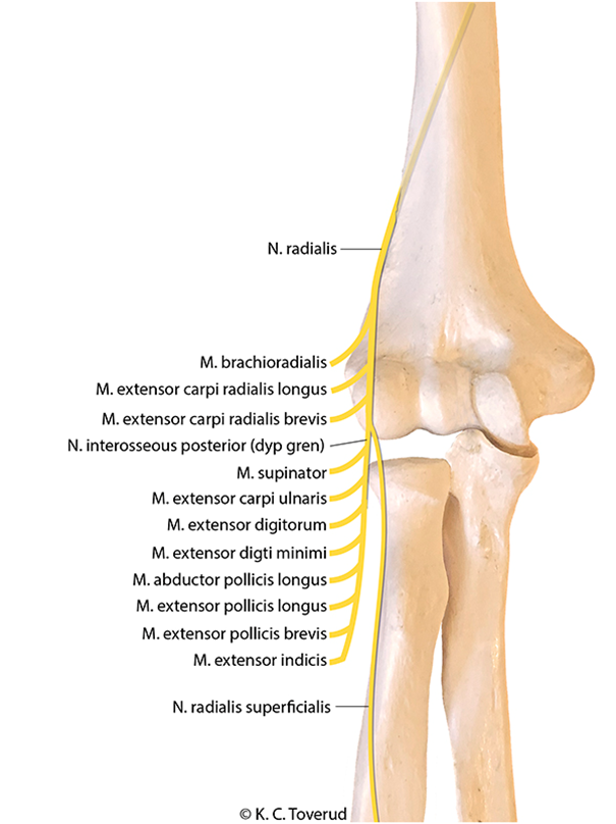
- Normal supination strength; innervation to M. supinator branches off before Frohse's arcade.
- Normal strength in radial extension; innervation to M. extensor carpi radialis longus and M. extensor carpi radialis brevis branches off before Frohse's arcade.
- No hand drop.
- When attempting wrist extension, there is a radial deviation of the hand due to weak M. extensor carpi ulnaris, innervation to M. extensor carpi ulnaris arises in Frohse’s canal
- Weakness in finger and thumb MCP extension, thumb IP extension, and thumb abduction
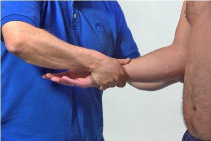
Posterior interosseous nerve syndrome, Supination test
Isometric supination increases the pain
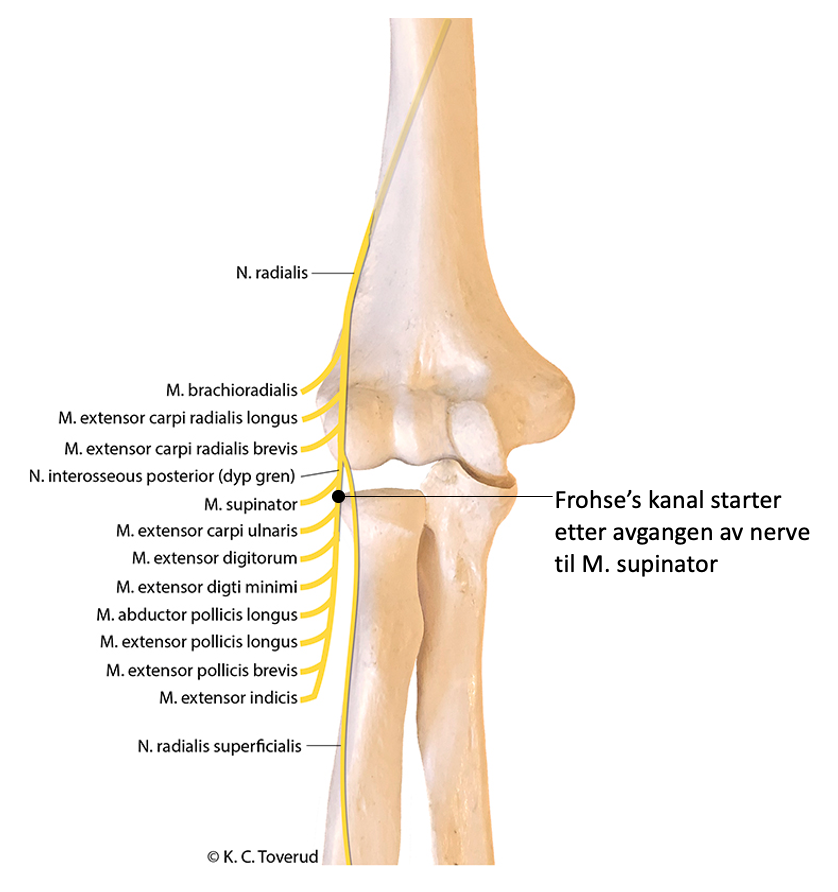
Key diagnostics, N. radialis