
Clinical suspicion of ankle fracture
- Severe inversion or eversion trauma
- Visible misalignment
- Cannot bear weight
- Need for further clarification with X-ray according to Ottawa Rules
Mechanism of ankle fracture
- Inversion trauma
- Eversion trauma
- The movement of the talus inside the ankle mortise during the trauma determines the extent of injury we get in the fibula, tibia, and syndesmosis
- Type of fracture injury is classified as Weber type A, B, and C
- Weber type A: Adduction force on supinated foot
- Weber type B: External rotation force on supinated foot
- Weber type C: External rotation force on pronated foot
Weber type A, 20-25% of fracture injuries, Adduction force on supinated foot
Weber A with moderate adduction force. Transverse fracture (avulsion fracture) in the lateral malleolus below the syndesmosis level. The tibiofibular syndesmosis is intact.
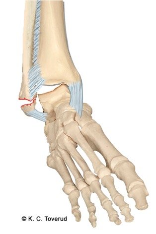
Weber A with large adduction force. Transverse fracture (avulsion fracture) in the lateral malleolus below the syndesmosis level in combination with oblique fracture in the medial malleolus. The tibiofibular syndesmosis and deltoid ligament are intact. Unstable fracture.
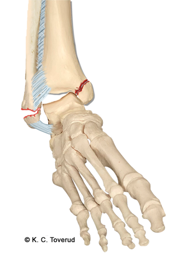
Weber type B, 60% of fractures, External rotation force on supinated foot
Weber B with moderate external rotation force. Oblique fracture of the lateral malleolus. The tibiofibular syndesmosis is injured.
Weber B with high external rotation force. Oblique fracture of the lateral malleolus, fracture of the medial malleolus, and fracture of the posterior malleolus. The tibiofibular syndesmosis and deltoid ligament are injured. Increased distance (fork gap) between the talus and malleoli. Unstable fracture.
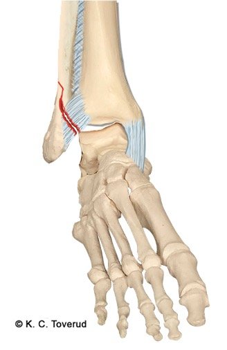

Weber type C, 20% of fractures, External rotation force on pronated foot
Weber C with moderate external rotation force. Transverse fracture of the medial malleolus with injury to the deltoid ligament and minor injury to the tibiofibular syndesmosis

Weber C with high external rotation force. Transverse fracture of the medial malleolus with injury to the deltoid ligament and injury to the tibiofibular syndesmosis. High fracture in the fibula above the syndesmosis level. Possibly in addition, avulsion fracture of the posterior malleolus. Increased distance between the distal part of the tibia and fibula due to severe injury to the tibiofibular syndesmosis. Unstable fracture.
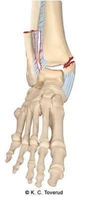
"Ottawa Rules", palpation suspected ankle fracture
Lateral malleolus, 6 cm proximal
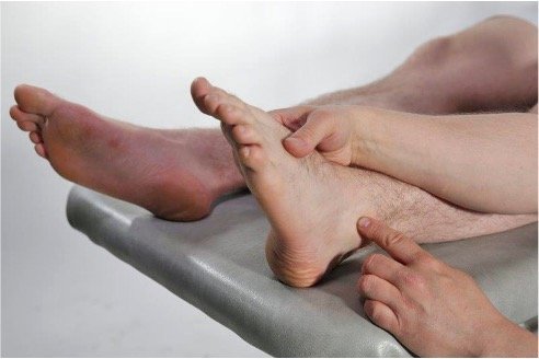
Medial malleolus, 6 cm proximal
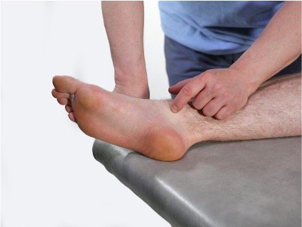
Base of 5th metatarsal
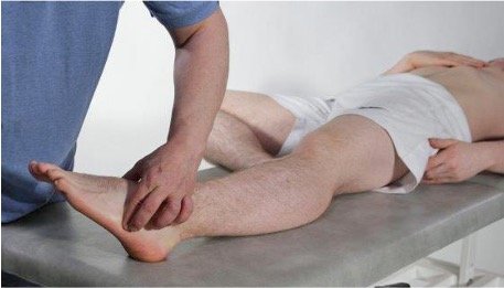
Navicular bone
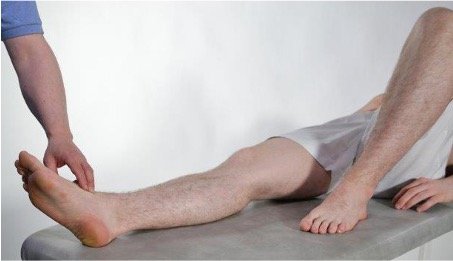
Ottawa Ankle Rules

- Need for X-ray of ankle when
- Tenderness of lateral malleolus
or - Tenderness of medial malleolus
or - Cannot walk four steps
- X-ray of foot when pain in navicular bone or base of 5th metatarsal or cannot walk four steps
- The rules are validated for individuals over 18 years, but can also be used as important guidance for younger individuals